Developments in Laboratory Scale Lyophilisation for Purification Laboratories

By: Dr Induka Abeysena & Rob Darrington

Introduction

For many years lyophilisation, or freeze drying, has been used to dry samples in the laboratory. The technique is well researched and has become the method of choice for many researchers with a few samples to dry. Lyophilisation is often preferred because a high level of dryness is achieved with low residual solvent levels, and because of the light, powdery, ‘fluffy’ finish of the sample which enables the sample to be easily removed and weighted out.
 
However, there are a number of potential drawbacks encountered with the conventional freezedrying apparatus, these include:
1.     Samples must be prepared in a limited range of solvents, normally only water can be used
2.     Volatile organic solvents and their mixtures cannot be used
3.     The process is slow

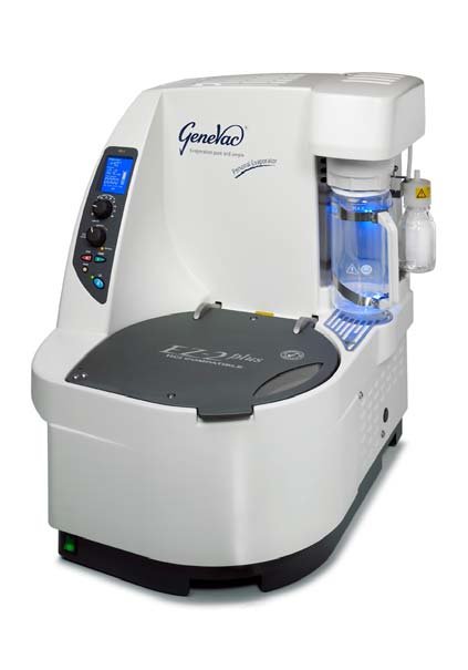
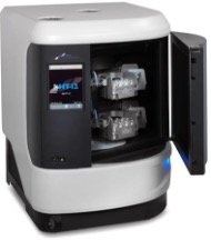
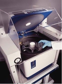
Therefore researchers with many samples to process, or mixtures of solvents, say from preparative reverse phase HPLC separation containing water and acetonitrile, have turned to centrifugal evaporators. In other laboratories, compound handling for example, the aggressive nature of the organic solvents used renders a freeze drier unsuitable. Even in for these environments, state of the art centrifugal evaporators, such as the Genevac HT-4X, shown in Figure 1, have some limitations. Problems reported include; samples are dried to a film and therefore may be difficult to resuspend after drying, some samples trap a little residual solvent, and some samples fail to dry with the majority.

Figure 1
Genevac HT-4X Centrifugal Evaporator

In this study we will present the results of research done in the Genevac laboratory to develop a ‘best ofboth worlds’ solution, where rapid parallel drying can be achieved while providing high levels of dryness, the ‘fluffy’ finish desired by many, and where every sample driesevery time.  

Problems with Purification

Within purification laboratories samples are typically presented dissolved in water and acetonitrile, with a low level of a modifier present, normally 0.1% TFA. Using a freeze drier to remove these solvents is fraught with difficulties, firstly, the acetonitrile requires a very deep vacuum to freeze it, or a freeze drier which actively freezes the samples. Acetonitrile freezes at –65°C. If the acetonitrile is not frozen thenbumping is inevitable resulting in sample loss and cross contamination.

Secondly, acteonitrile in the ice trap will spoil the vacuum making lyophilisation of the water almost impossible. Thirdly, the process is slow, which is incompatible with the drive to reduce process times within many industries. For these reasons the centrifugal evaporator has become the method of choice, because it issuited to rapidly drying may samples in parallel, and designed to control bumping when drying solventmixtures. 

However, there are two potential problems, both are sample related effects. Users report that they experience difficulties with drying a few samples per batch. The problems may be that not all the TFA is removed, residual TFA may damage the sample when in storage, and secondly, the compound may interact with the water boosting the boiling point, making drying difficult. Additionally, residual solvent shows up in Nuclear Magnetic Resonance (NMR) analysis. Whilst these problems occur occasionally, the implications of picking a few samples by hand are prohibitive for many automated laboratories, thereforethe whole sample rack is reprocessed. 

Lyophilisation in a Centrifugal Evaporator

Samples prepared in water can be lyophilised in a centrifugal evaporator by pulling the best vacuum available, in the Genevac HT-4X (Figure 1) fitted with the solvent resistant scroll pump, the ultimate vacuum is well below 0.5mbar which is more than adequate to freeze water. This process is akin to normal freeze drying and therefore slow. 

Genevac developed a process some years ago where by users can evaporate some of the solvent using all the speed of a centrifugal evaporator, and then switch to a lyophilisation mode when only a few millilitres of solvent are left, thus delivering the best of both worlds. For one Genevac customer this took process time for 96 x 30ml fractions from 48 hours in a freeze drier, to 16 hours (an overnight process) in a Genevac HT-12, equivalent to only 10 minutes per 30ml sample, were the samples dried sequentially. Using this as a platform, the effects of heating a sample during lyophilisation were studied to determine if this gave a speedadvantage. 

The study of water containing samples was in two halves. Initially just water was used to develop the optimum conditions, and then with water and acetonitrile to simulate samples taken from HPLC.

Lyophilisation of Water

In all trials Ibuprofen sodium salt was used as the standard sample. A stock solution of 0.01M was prepared in water. 15mls of solution was loaded into each of 48 20ml scintillation vials (Wheaton) and placed dried in a Genevac HT-4X evaporator under various conditions.  Figure 2 shows a typical vial holder.

Figure 2
Genevac 20ml scintillation vial holder.

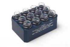
Figure 3 shows a plot of concentration and then lyophilisation of water, this was developed to establish a baseline. The settings used are summarised in Figure 4. The total time taken to dry the sample is approximately 8 hours, 4 hours of concentration in stage 1 & 2, where the sample temperature is at about +8°C and then stage 2, the lyophilisation stage where the sample is frozen to –16°C and warms up whendry.

Figure 3
Baseline method showing concentration and lyophilisation of water based sample – Trial 1

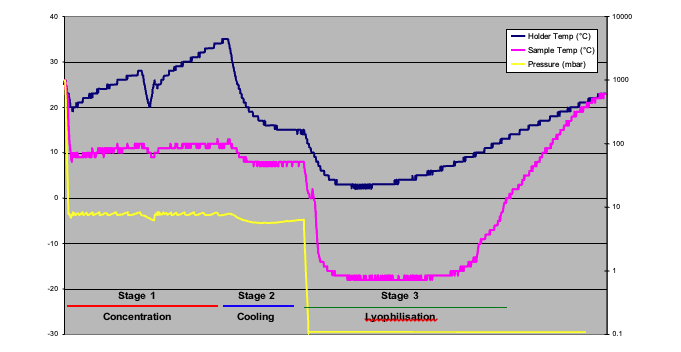
Whilst there are three stages shown in Figure 3, the actual evaporation method comprises up to four stages:

1.     Concentration of the bulk of the solvent using fast evaporation

2.     Cooling of the samples and sample holders, in preparation for

3.     Freezing of the sample using deep vacuum

4.     Lyophilising the residual solvent, with or without heat

Figure 4
Results of Lyophilisation trials with water

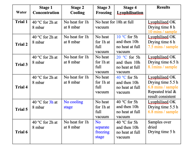
Figure 4 shows the results of the method development for the water processing stage. During sequential runs different heating levels were used in the lyophilisation stage, higher heat levels were shown to reduce the total processing time from 8 hours in trial 1, to 5.5 hours in trial 4. Figure 5 shows the results of trial 4.

The precise processing time for each trial was not known, therefore each sample was over processed, and then from the data the end point was identified. For the sake of uniformity, the time at which the sample temperature became positive was taken as the end point. At this time the samples are warming up rapidly, because there is no longer any cooling effect from lyophilisation.

Figure 5 – Results of concentration and lyophilisation with heating, Trial 4

Points to note from these data – we had thought that best practice demanded a cooling stage, stage 2, this had always worked in the past, but had never been tested. In effect the freezing stage achieves cooling as well as freezing, therefore this stage is not necessary, as demonstrated by trial 5. However, the freezing stage with no heat appears to be essential, as shown by trial 6, in this case the samples dried normally, and did not lyophilise, it was evident from the data that the sample had not frozen at all. The results of trial 3 appear to be anomalous, in that higher heat should reduce the processing time, but does not in this trial.

Lyophilisation of Water & Acetonitrile

For many users lyophilisation of water is trivial, whilst the time savings demonstrated in our study are welcome, the issue remains of how to deal with solvent mixtures. A modification to the method used for water is the addition of an earlier stage, stage 0, to remove the acetonitrile before concentration of the water.
 
Stage 0 has three parts:
1.     Dri-PureTM – vacuum ramping and high rotor speed to prevent bumping
2.     Concentration – a 40mbar stage to remove the acetonitrile without freezing the water, at 40mbaracetonitrile boils at +2°C
3.     Draining the condenser – residual acetonitrile will spoil the vacuum in later stages, therefore must be removed.

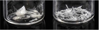
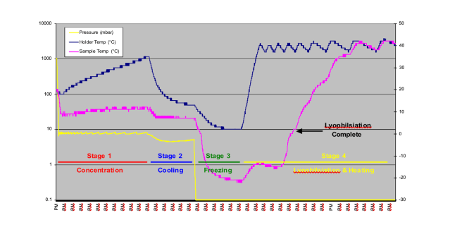
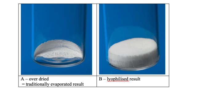
For these trials a 0.01M solution of Ibuprofen sodium salt was prepared in a 60:40 mixture of water and acetonitrile. Figure 6summarises the results. As is evident from the data, it was only at trial 10 that the optimum conditions were achieved. As withthe water trials, the cooling stage was not required.           The process needed adjustment to achieve the correct balance of concentration and lyophilisation. 48 x 15ml samples dried in 5 hours, equivalent to 6.25 minutes if the samples were driedsequentially. Figure 7 shows the difference between a lyophilised result, achieved in trial 10, and traditionally centrifugally evaporated sample as per trials 7, 8, 9 and 11. The difference is stark, and the ease of resuspension is greater with the lyophilisedsample, where as the dried sample does not fully dissolve readily. The resuspension of the samples can be viewed at http://www.genevac.com/products/lyophilisation.html

Figure 6
Results of Lyophilisation trials with Water and Acetonitrile
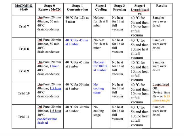
Figure 7
Dried samples in Scintillation vials

Conclusions & Discussion

Lyophilisation of the samples improved the ease of redissolving the sample post drying. The addition of heat during the lyophilisation stage reduced the lyophilisation time considerably. When establishing the lyophilisation method, the traditional cooling stage is not required, but the freezing stage has been shown to be essential.
When evaporating water and acetonitrile mixtures it is necessary to drain the condenser following evaporation of the acetonitrile and before evaporation and lyophilisation of the water. Residual acetonitrile in the condenser spoils the vacuum preventing lyophilisation conditions being achieved.
 
Using the HT-4X the system had to be drained manually at the end of stage 0, some Genevac systems are able to automate this facility eliminating the need for user intervention mid process. An option to automatethis on the Genevac HT-4X and HT- 12 is being developed as a result of this work.

Evaluation of Evaporative Sample Preparation Techniques for the Extraction of Drugs of Abuse from Urine Samples by Forensic Science Ireland.

By: Paula Clarke, Forensic Science Ireland. Alison Wake, Genevac Ltd.

Introduction

A method for the extraction of drugs of abuse from urine samples pertaining to drug facilitated sexual assault cases has been developed in house by Forensic Science Ireland (FSI). The method involves the use of solid phase extraction techniques prior to screening analysis by LCMSMS. As part of the solid phase extraction  process, the extracted sample eluent is evaporated to dryness. FSI evaluated the use of a Genevac EZ‐2       personal evaporator (Figure 1) as an alternative method of evaporation as part of their validation process.

The EZ‐2 was chosen due the fact that it eliminated the need for gases such as nitrogen, required for the       conventional blow down evaporation method, and also reduced sample handling and reformatting stages.

Sample preparation method

Urine samples (1ml) are first hydrolysed by incubation with 500ul ß‐glucuronidase enzyme (prepared in 0.1M acetate buffer) for 1ó to 2hr at 60C. 100ul of deuterated internal standard is also added. Following centrifugation at 13,000rpm for 2min the supernatant is removed and 1ml borate buffer added to adjust the pH to ~9.

SPE extraction is carried out using a 3cc 60mg SPE cartridge as follows:

  • Condition ‐ 3ml MeOH
  • Equilibrate ‐ 3ml Water
  • Load sample
  • Wash ‐ 3ml 5%MeOH
  • Max pressure 50psi for 5 mins to dry bed
  • Turn off Flow Rate
  • Elute ‐1ml MeOH
  • Elute ‐ 1ml 3:1 MeOH:IPA
  • Max pressure to drive off remaining eluent

The resultant 6ml of eluent must then be evaporated to dryness and reconstituted in 200ul diluent prior to analysis by LCMSMS.

Choice of Evaporation Procedure

Under the conventional method, evaporation was achieved using a nitrogen blow down system. Evaporation of the eluent by blow down in the SPE fraction collection tube results in the analytes being deposited up the sides of the tube. With a reconstitution volume of only 200ul, it is difficult to ensure that all the analytes have been resuspended. To ensure maximum recovery, evaporation needed to carried out in the MS analysis vial; the 6ml of eluent being added in 2ml aliquots and evaporated to dryness in three stages.

The EZ‐2 (Figure 1) is a centrifugal evaporation system, which works by boiling solvent at low temperature under vacuum.

During evaporation the samples are centrifuged at around 500G primarily to prevent bumping and subsequent cross contamination between the multiple samples which are being dried in parallel. Centrifugation also results in the dried analyte being deposited at the base of the tube. Reconstitution in very low volumes is therefore possible. Using the EZ‐2, the full 6ml of eluent sample can be dried and subsequently reconstituted to 200ul diluent directly in the SPE fraction collection tube.

The EZ2 was evaluated in terms of its ability in achieving required limits of detection, sample integrity and the risk of cross contamination.

Limit of Detection Study

The FSI urine screening method by LCMSMS covers a wide range of drugs of abuse. UNODC’s (United Nations  Office on Drugs and Crime) Minimum Required Performance Limit (MRPL) were achieved for all drugs /  metabolites listed in the FSI screening panel using the EZ2. Studies performed by FSI showed that increased  sensitivity was achieved by using the Genevac EZ2 evaporator compared to other conventional evaporation  techniques. For example at the recommended UNODC MRPL, greater sensitivity was achieved when using the EZ2 for 7‐Aminoflunitrazepam, Morphine, Amphetamine and Phenazepam among others, when compared other common methods of evaporation. 

Sample Integrity Study

A parallel study was performed using the conventional blow down method versus the EZ2 as a means of  evaporation. The conventional process involved evaporation at a temperature of 45°C whereas the use of the  EZ2 allowed evaporation at a lower temperature of 40° C to be achieved with sample integrity being  maintained. Additionally, the time taken to complete the evaporation process was halved by using the EZ2.

Cross Contamination Study

The aim of the cross contamination study was to determine if any cross contamination occurred during the evaporation process in the Genevac EZ2 for common targeted drugs of abuse in urine samples related to drug facilitated sexual assaults.

The study involved using the EZ2 to evaporate to dryness a series of spiked urine samples, which had undergone solid phase extraction. Each spiked urine sample contained 43 various drugs of abuse / metabolites at a concentration of 500ng/ml in addition to 6 deuterated internal standards*.

The extracted sample eluent from the spiked urine sample was held in a 6ml fraction collection tube and was placed in the sample holder surrounded by fraction collection tubes of blank eluent – known as the ‘blank sample’. All samples (spiked urine and blanks samples) were evaporated to dryness in the EZ‐2, reconstituted in diluent and analysed by LCMSMS.

For the spiked urine samples all drugs / metabolites were detected. All the ‘blank’ tubes, surrounding the spiked urine samples were negative indicating that cross contamination did not occur during the evaporation process using the EZ2.

Conclusions

The experiments performed by FSI have all indicated that evaporation using the EZ2 results in a more streamlined process. Less time is required for completion of the evaporation process and sample integrity is maintained. Increased sensitivity was also achieved without the risk of cross contamination – ultimately a more superior process to conventional methods of evaporation. The method has been accepted into routine use.

*Drugs / Metabolites and Deuterated Internal Standards contained in the spiked urine sample:

Chlordiazepoxide, Flurazepam, Zopiclone, MBDB, Diamorphine, Oxycodone, Norbuprenorphine, Temazepam, Midazolam, Oxazepam,  Clobazam, MDEA, Hydromorphone, Dihydrocodeine, Methamphetamine, Methadone, Morphine, THC, THC‐COOH, 7‐  Aminoflunitrazepam, Clonazepam, Nitrazepam, Cocaine, Diazepam, Alprazolam, Lignocaine, MDMA, Amitriptyline, Codeine,  Nordiazepam, Lorazepam, Phenacetin, Amphetamine, Zolpidem, Buprenorphine, Bromazepam, 6‐Monoacetylmorphine, MDA,  Phenazepam, Ketamine, Triazolam, Flunitrazepam, Benzoylecgonine, Cocaine‐d3, Codeine‐d6, Amphetamine‐d11, THC‐d3, Diazepam-d5,  Methadone‐d9

Evaluation of an Improved Sample Preparation Method for Quantative Analysis of Very Low Levels of Airborne Polycyclic Aromatic Hydrocarbons for Worker Protection and Health Screening

By: Nicolas Falquet, Gilles d’Esperonnat & Rob Darrington

Introduction

Polycyclic Aromatic Hydrocarbons (PAHs) are large class of compounds comprising two or more fused aromatic rings. PAHs are naturally occurring in fossil fuels and their derived products and can be formed during incomplete combustion of carbon based fuels. As such they are a by-product of many industrial processes. PAHs vary greatly in size, nature and hazard to human health, some are not classified as toxic, where as others are known carcinogens. The IARC specified 16 as being of particular interest, others have subsequently added this list. In all, over 100 PAHs have been described.

Given the risks and potential risks to human health presented by PAHs, many high risk organisations, such as Foundries, Bitumen Works & Smoke Houses routinely monitor workers and their environment for PAH levels. Typically PAHs are trapped using filters (particulate forms) or resins such as XAD2 (gaseous forms) through which work place environmental air is drawn. Filters may be situated in a small device attached to the workers overalls, or from larger units measuring the air in a wider area. Potential problems exist when recovering the PAHs from the filters and preparing the samples for analysis, principally, losses due to PAH volatility are reported for bi- and tri-cyclic PAHs (ISO11338-2:2003). Therefore, ITGA undertook a study to improve sample recovery and therefore PAH determination when working with low and very low levels of analytes.

Sample Preparation Methodology

Methods for workplace sampling are well described in the literature (NFX43-294 and Method Metropol 011) and result in samples trapped on glass or quartz fibre filters. The filters are preserved and delivered to the analytical laboratory. The whole filter placed into a barcoded vial, 10ml dichloromethane (DCM) is added and the tube placed in an ultrasonic bath at room temperature for 15 minutes to extract the analytes. This operation is repeated once with 10ml of DCM to optimise extraction. Following extraction the sample is concentrated to 1ml using a nitrogen blowing system and then analysed via HPLC coupled to a Fluorescence detector. XAD2 resin tubes may be used as an alternative to fibre filters.

Evaluation of new Sample Preparation Methodology

A standard solution containing the US-EPA 16 PAHs (as defined by IARC, 1987) was spiked onto quartz fibre filters or XAD2 resin tubes and allowed to air dry. The filters / tubes were then extracted twice using 7ml DCM and sonnication in the ultrasonic bath for 15 minutes at room temperature. The combined sample (14ml) had a 100ul aliquot removed. This was made up to 1ml with acetonitrile was taken and injected into HPLC-Fluorescence to provide a 100% reference. The remaining DCM had 100l 2-pentanol added as a solvent keep and was evaporated via centrifugal vacuum evaporation in the Genevac EZ-2 Envi (Figure 1). Temperature and pressure during evaporation were controlled such that the DCM evaporates but the 2-pentanol does not, as previously described by Marsico (2006) and Massat et al. (2007).

Figure 1 (right) – Genevac EZ-2 Envi

The samples were then made up to 1ml using acetonitrile and injected into HPLCFluorescence for analysis. Recoveries for all analytes, even the most volatile were in excess of 90% and the fit of the analytical curve to the reference sample was very good, and shown in figure 2 below.

The samples were then made up to 1ml using acetonitrile and injected into HPLCFluorescence for analysis. Recoveries for all analytes, even the most volatile were in excess of 90% and the fit of the analytical curve to the reference sample was very good, and shown in figure 2 below.

Figure 2 – HPLC-Fluorescence Chromatogram Overlay of Reference Sample to Post Concentration Sample

Red – the reference point. Blue – other chromatograms refer to the PAH compounds Naphthalene, Acenaphthene, Fluorene, Phenanthrene

Validation of the Process

Having delivered similar results to the existing method, and being beneficial in the sense of “automation” of the concentration process, statistical validation of the process and equipment was required. Using the above methodology, a solution containing 14 PAH samples was spiked onto quartz fibre filters and also on to XAD2 resin tubes. Filters were spiked at 100ng and 10ng. These were allowed to dry and extracted, concentrated and analysed. The process was repeated on six distinct occasions using new samples and solutions on each occasion. The results are presented in Figure 3.

Figure 3 – Data from Validation Studies 

Mass Recovered (ng) and Recovery % are averages from each of the 6 repetitions performed. SD is the standard deviation across repetitions.

The results generally show excellent recovery and good standard deviation figures. Due to a contamination from XAD2 resin, for two compounds (naphthalene and acenaphtene) limits of quantification have been validated at 50ng instead of 10ng.

Conclusions

The new method of sample preparation was found to be superior to the existing methods. Recoveries are seemingly a little lower for the 10ng studies because this  approaches the limit of detection of the analytical method. Following successful validation and external audit by COFRAC (Comité français d’accréditation) the new method and systems have been adopted into routine daily use.

About the Authors

Nicolas Falquet is Testing Manager at ITGA, a leading independent analytical testing laboratory, based at Le polygone, 46 rue de la Télèmatique, 42000 St-Etienne, France.  ITGA is part of the Carso Group.

Gilles d’Esperonnat is responsible for sales and service of Genevac evaporators in France and based in the Lyon area Rob Darrington is Product Manager at the Genevac head office, Farthing Road, Ipswich, IP1 5AP, UK.

References

IARC. 1987. IARC Monographs on the evaluation of carcinogenic risks to humans, supplement 7, Overall evaluation of carcinogenicity: an updating of IARC monographs 1-42. Lyon: International Agency for Research on Cancer

Marsico, Anna Maria, 2006. Improving Analysis of Pesticides – a new method development protocol to increase recovery of volatile compounds. First published in Lab Asia, August 2006 & available via  http://www.genevac.org/en/ArticleDetail.asp?S=6&V=1&ProductDownload=81

Massat, F, Planel, B & Venezia, A, 2007, Evaluation of Evaporative Sample Preparation Techniques. First published in International Environmental Technology, March/April 2008, pp 36, and also available via http://genevac.org/en/ArticleDetail.asp?S=6&V=1&ProductDownload=134

NF X 43-294. June 1995. Sampling and analysis of polycyclic aromatic hydrocarbons INRS. 2007. Method Metropol 011. Polycyclic Aromatic Hydrocarbons. NF ISO 11338-2. March 2004. Determination of gas and particle-phase polycyclic aromatic hydrocarbons – Part 2 : sample preparation, clean-up and determination.

An Improved Evaporative Sample Preparation Methodology for Determining Nitrofuran Antibiotic Residues in Foodstuffs

By: A. Kaufmann; K. Maden and S. Walker

Introduction

Nitrofuran antibiotics were banned from use in the European Union [EU] in 1995 due to concerns that their residues were carcinogenic. In 2002/3 the EU introduced a stringent testing regimen which calls for the use of highly sensitive methods to test food stuffs, principally meat, fish & shellfish, for the presence of this class of antibiotics. The Minimum Required Performance Limit [MRPL] laid down by the EU directive is 1g per kg for edible tissues, and is enforced on all products whether produced locally or imported into the EU. Many papers detail methods and identify metabolites and derivatives of the drugs concerned and are listed by Vass,

Hruska & Franek (2008). The analytical method calls for good upstream sample preparation to eliminate the effects of the matrix, and can be manual and time consuming, particularly where evaporation is concerned. This article describes operational benefits including workflow improvements gained by the official food control authority of the canton of Zurich (Kantonales Labor Zurich) or KLZH during improvement of their upstream sample preparation methodology.

Sample Preparation Methodology

Of the methods cited in the literature for upstream sample preparation, many laboratories favour physico-chemical assays with chromatographic separation and mass spectrometry [MS] detection of metabolites.

The general scheme for preparation and analysis of nitrofurans in foodstuffs at KLZH is as follows, and summarised in figure 1:

1. Homogenisation of tissue sample

2. Acid hydrolysis to release tissue bound metabolites

3. Derivatisation of metabolites with orthonitrobenzaldehyde to increase molar mass and increase sensitivity of detection

4. Liquid:liquid extaction with ethyl acetate.

5. Evaporation to dryness.

6. Resuspension with water.

7. Clean-up with Solid Phase Extraction to eliminate interfering matrix elements, such as lipids

8. Elution with ammoniacal methanol.

9. Evaporation to dryness

10. Resuspension in a known small volume of solvent suitable for chromatography.

11. Separation via ultra high performance liquid chromatography [UHPLC] and analysis with MS/MS.

The most time consuming and labour intensive parts of this methodology are the extraction and the two evaporation-to-dryness steps.

Traditional Evaporation Technique

At KLZH the evaporation part of the sample preparation method has traditionally relied upon use of a rotary evaporator. The rotary evaporator method for Nitrofuran testing was proven to give good recoveries. However it has a number of operational drawbacks. The biggest disadvantage was that the rotary evaporator is a single sample system which requires continuous monitoring to control the process and to ensure that no foaming or bumping occurs. Samples for Nitrofuran typically presented to KLZH in batches of 5 to 30. Where a small batch is managed in an assessable amount of time with a rotary evaporator, bigger batches would soon become impossible to process. Therefore, three batch process evaporators were evaluated in order to improve the productivity of this step.

New Evaporative Technologies Evaluated

Blow-Down Evaporation
In these evaporator systems, an inert gas such as nitrogen is blown down through needles onto the samples in tubes to create a flow over the liquid surface. This alters the equilibrium between the vapour and liquid phases to favour the vapour phase. Heat is normally applied to the samples to hasten evaporation. Therefore the samples are hot during the process, being at the temperature of the heating block or bath, and consequently the technique offers poor recovery of volatile analytes. Although blow down evaporation is relatively fast for volatile solvents, it can be slow for solvents with high boiling points or those that are difficult to evaporate such as water. Blow down evaporation requires continuous monitoring by the user to detect the end point of the drying process and to optimise the position of the gas jets, keeping them close to the liquid surface.

Vortex Evaporation
These systems boil batches of samples under vacuum and therefore the samples are cold throughout evaporation, while swirling the sample tubes to create a vortex. The vortex created generates a large sample surface area for evaporation, making the process relatively fast.

However, the resultant dried product is spread across the vessel walls, which can make sample recovery more difficult. Moreover, in contrast to centrifugal concentrators, the swirling movement generates insufficient g force to prevent solvent bumping and tends to aid foam formation. Hence vortex evaporators are prone to sample loss and cross contamination. Controls on the system can permit hands free operation once the vigour of the vortexing action has been correctly set.

Centrifugal Vacuum Evaporation
Centrifugal evaporators induce solvent boiling under vacuum and so the samples are cold. Centrifugal evaporators use cold traps to recover the vaporised solvent. Centrifugation ensures that solvent boils from the sample surface downwards, thereby eliminating boiling over, foaming and solvent bumping and so preventing sample loss and cross-contamination. Solvent at the liquid surface is at the pressure of the equipment, whereas solvent below this level is at a higher pressure due to the extra weight of solvent multiplied by the g force exerted by the centrifuge rotor. Systems with very high rotor speeds generating 500g or more are proven to prevent solvent bumping. The centrifugal evaporation technique accommodates a wide range of solvents and can concentrate, dry to a film or freeze dry samples. Controls on the system permit hands free operation, with the most advanced systems having automatic detection for the end of the method built in.

Results of Evaluation

The systems were tested for their suitability for use in the sample preparation process, with particular attention to cross-contamination / bumping / foaming, solvent recovery and degree of user intervention. The results are shown below in Figure 2.

Figure 2 (Below) – Results of solvent evaporation system evaluation

Conclusions

Working with the rotary evaporator, a batch consisting of a total of 20 samples (reference solutions, samples and spikes) would require approximately 1.5 hours (evaporation time) for the first evaporation step and another 2.5 – 3 hours for the second evaporation. A total of 4 – 4.5 hours spent where the operator has to keep an eye on the evaporator and change the samples every few minutes.

The centrifugal evaporator would require for the same batch up to 3 hours for both evaporation steps. In that time, the operator is free to do other work.

The centrifugal evaporator can take a total of 48 nitrofuran samples. These 48 samples would require approximately one hour more to evaporate (both steps) than a batch of 20.

This would be an estimated 2 – 3 hours saved in comparison to working with a rotary evaporator.

Overall the installation of the EZ-2 Envi evaporator (Genevac Ltd, Ipswich, UK. Shown in figure 3) at KLZH is estimated to save the laboratory 2-3 hours per day that previously was spent on evaporation tasks.

The instrument enabled the processing of larger sample series within a given day. It significantly reduced the contamination (carry-over) issue. Previously, the frequent false positive findings were the reason for re-analysis of affected sample or whole sample series.

Very important is also the fact that the EZ-2 Envi operates unattended in a fully automatic manner.

References

Vass, Hruska & Franek. 2008. Nitrofuran Antibiotics: a review on the application, prohibition and residual analysis. Veterinarni Medicina, 53, 2008 (9), pp 269-500.

About the Authors

A. Kaufmann, K. Maden und S. Walker are working at the official food control authority of the canton of Zurich (Kantonales Labor Zurich). The group focuses on the analysis of veterinary drug residues in animal based food products. Commonly uses analytical techniques are Liquid chromatography coupled to high resolution mass spectrometry and liquid chromatography coupled to tandem quadrupole mass spectrometry.

Improved Stability Drug Development

By: Ian Bailey, Biopharma Process Systems

Background: Why there is a need for the Exalt™ application

Polymorph screening is an important stage of drug development. The aim is to identify the different crystalline structures or polymorphs that a drug may appropriate. The information gained is used to optimise the physical properties of the drug compound to ensure efficacy, and provide formulation and manufacturing consistency.

Ibuprofen crystallised from Acetone (left), and Ethyl Acetate, (right)

Once a drug compound is discovered, consideration must be given on how it will be administered to a patient. A drug compound may have poor water solubility, poor stability or a lack of crystalline form. This is usually overcomeby creating a ‘salt’ version of the drug compound.
Compounds that are crystalline in nature, often adopt a number of crystalline forms or polymorphs. Different polymorphs will have different physical characteristics which can impact on the manufacturing process, which canaffect the efficacy of the drug.

In the case of the anti-viral drug Ritonavir, not only was one polymorph virtually inactive compared to the alternative crystal form, but it was subsequently found to convert the active polymorph into the inactive form on contact due to its lower energy and greater stability, making spontaneous interconversion energetically favourable. Even a speck of the lower energy polymorph could convert large quantities of Ritonavir into the inactive polymorph, and this caused major production issues until   the drug was finally reformulated.

It is also essential for all crystal forms to be listed on any drug patent. In the case of Prozac, an Eli Lilly drug, a generic drug manufacturer discovered another form not listed on the patent, and subsequently released a copy of the drug. A year prior to patent expiry, Eli Lilly lost several billion dollars in revenue as  a result.

Regulatory authorities expect drug manufacturers to conduct thorough assessments of new drugs to form polymorphs. Pharmaceutical companies address the issues by carrying out a screen on the chosen compound to identify as many different polymorphic forms as possible. Ideally, this screen takes place at an early stage in the drug development cycle so that a manufacturing process can be defined to produce the correct form for clinical trials and the pharmaceutical market. A rational and extensive initial screen will be repeated as necessary throughout the project, for example, if the impurity profile of the material changes, the robustness of the crystallisation process will also be thoroughly investigated.

The screening process is time and resource consuming with no guarantee that all polymorphs will be discovered. It can also require a significant amount of active pharmaceutical ingredient (API). Polymorph studies are carried out manually, and each chemist usually employs slightly different approaches which can result in a lack of consistency.

Screening for polymorphs involves applying a wide range of crystallisation conditions in a number of solvents with diverse properties, (eg; polar, non-polar, aromatic etc). Applying different conditions to the materials in the solid phase may also generate new forms. Of particularimportance is the variation of the rate of crystallisation – slower crystallisation typically yields thermodynamically stable forms, whereas faster experiments will produce metastable forms that may be kinetically preferred. When new forms are identified, they may, in turn, be used asinput material for further experiments.

Studies for polymorph screening can therefore take a long time and may be difficult to conduct. Working with researchers in the field, Genevac Ltd has developed Exalt™ (Figure 1.), a unique toolkit to help researchers conduct evaporative crystallisation studies in a number of ways, whether polymorph screening, or searching for metastable and stable forms. Many crystallisation tools are currently available, and researchers usually use all ofthem in an effort to reduce the risk of new forms appearing later in the project. Although these techniques cannot all be replaced by Exalt™, most, if not all of theevaporative methods can be, so the crystalline forms of a drug can now be delivered in a controlled andreproducible manner.

The development of Exalt™ by Genevac Ltd, began with a customer request to utilise evaporation in a controlled way to produce stable crystal forms, ie; very long evaporation times, (>72 hrs), evaporation of a wide range of solvents at the same time and at the same slow rate of evaporation for every solvent, volatile through to non-volatile. Analysis by X-ray diffraction (XRD) and Thermogravimetric differential thermal analysis, (TG-DTA) confirmed the presence of truepolymorphism rather than solvation.

How does Exalt work?

The Exalt™ software allows the pressure reduction from atmospheric and the cycle time to be carefullycontrolled so that evaporation can take place from 6 hrs to 72 hours or more. The condenser must be able todefrost automatically every 6 hours, or a large plug of ice forms in the condenser inlet.

The combination of pressure cycling and baffles delivers slow, controlled evaporation of different solvents at the same time and at similar rates. Studies of solvents with BP’s in the range 40⁰C to 165⁰C, DCM toDMAc (including water) have been made possible.

Pressure cycling creates a serial dilution of the atmosphere surrounding the samples. Spinning the rotor induces a vortex in the tower, promoting vapour diffusion. This “chimney” effect helps the non-volatile solvents. Bafflesslow the flow of vapour from one chamber to the next.

Towers containing baffles sit on top of the vials. The baffles have varying diameter holes, and are selected depending on the solvent properties. More baffles, with smaller holes for volatile solvents, fewer, (or no) baffles and larger holesfor non-volatiles.

Figure 2. Left to Right: Exalt baffles & tower assembly – Loading vials & towers – Complete holder assembly

The tower containing the baffles has four sections; base, which can have a seal and a baffle fitted, and  the remaining three top sections which can each have a baffle fitted.

The Exalt™ application will happily run on the new Genevac series 3 HT systems (Figure 3), or older series 2 HT systems fitted with auto- defrost and drain condensers, ICOPS controls and version 3.06 software or later, to give anadditional pressure control option, “crystallise”. This then enables the user to set the pressure reduction (from atmospheric) and the cycle time. The necessary upgrades are available on older series 2 HT systems, (depending on the age of the system, suitability can be checked by providing the manufacturer’s serial number).

During the summer of 2013, Researchers at Novartis Pharmaceuticals in Horsham, UK carried out a lengthyevaluation of eXalt™ technology to investigate how it may be applied to small molecule crystallisation processes in pharmaceutical chemistry R & D. Conclusions from this study were that Exalt offers a simple, reproducible method for evaporative crystallisation screening. The method is easy to use and allows late stage medicinal chemists to easily screen for crystalline forms with as little as 5mg of compound. Being non-destructive valuable compound is not lost and can be reused and knowledge gained can be used to develop a classic scalable crystallisation process. Crystals produced by exalt are high quality and suitable for XRPD analysis as well as for useas seed crystals.

FirstpublishedinManufacturingChemist,July2016

The Importance of Controlled Concentration and Drying in MALDI-TOF Applications

By: Steve Knight BSc Hons, MA, Marketing Manager, Genevac Ltd

Matrix-assisted laser desorption/ionisation-time of flight mass spectrometry (MALDI-TOF MS) is a now an accepted and routine analysis for the elucidation and quantitation of biomolecules in life science research. In this technique a co-precipitate of a UV-light absorbing matrix and a biomolecule are irradiated by a nanosecond laser pulse. The technique involves spotting small concentrated aliquots of material on to a matrix-coated “target”. The target is then positioned inside the Mass Spectrometer and the biomolecule of interest is desorped from the matrix surface and ionised by the laser. Most of the laser energy is absorbed by the matrix, which prevents unwanted fragmentation of the biomolecule, whilst some of the energy causes ionisation of the biomolecule. These ionized biomolecules are accelerated in an electric field and enter the flight tube of a time- of-flight mass spectrometer. During the flight in this tube, different molecules are separated according to their mass-to-charge ratio and reach the detector at different times. In this way each molecule yields a distinct signal. The method is used for detection and characterization of biomolecules, such as proteins, peptides, oligosaccharides and oligonucleotides, with molecular masses between 400 and 350,000 Da. It is a very sensitive method, which allows the detection of low (10-15 to 10-18   mole) quantities of sample with an accuracy of 0.1 – 0.01 %. Although the technique can be very sensitive, concentrated samples achieve the best results.

Protein identification by this technique has the advantage of short measuring times (a few minutes) and negligible sample consumption (less than 1 pmol) together with additional information on microheterogeneity (e.g. glycosylation) and the presence of by-products. Although molecular biology has provided powerful techniques for DNA analysis, this is not yet reflected in protein analysis. Genome sequencing has yielded a wealth of information on predicted gene products, but for the majority of the expressed proteins no function is known. Proteomics is an important new field of study of protein properties including expression levels, interactions and post-translational modifications and thus can be described as functional genomics at the protein level. The mass accuracy of MALDI-TOF MS is sufficient to characterise proteins (after tryptic digestion) from completely sequenced genomes such as methanogens and yeast. The use of MALDI-TOF MS for typing of single nucleotide polymorphisms using single nucleotide primer extension has also made important progress recently.

Oligonucleotides, proteins, antibodies and other larger biomolecules are all suited to MALDI-TOF analysis. However, these compounds can be difficult to concentrate without exposing them to thermal damage or cross-contamination. MALDI spotters use nano-scale liquid handling to pipette drops of sample on to the pre-coated target, but the sample is picked up from a well in a fairly standard microplate. The bulk sample is frequently formatted in a 96 well plate, with a number of plates contributing to each MALDI analysis run.

Concentrating large biomolecules in such microplates is not straightforward and it is here that Genevac’s centrifugal evaporation technology can help. By protecting samples from over-exposure to heat and by controlling cross-contamination in the plates, Genevac evaporators can significantly improve the results generated from MALDI-TOF analysis. This article looks at how that protection is achieved in practice.

Centrifugal evaporation is, of course, not new. The technique has been used in life science research for 20 years or more, but it was rather crude until fairly recently. Samples were spun sufficiently fast (it was thought) to hold the sample in the bottom of the container as it boiled (evaporated) whilst atmospheric pressure was reduced to induce boiling close to, or below, room temperature. To speed drying, heat could be applied by warming the chamber walls. More recently manufacturers added powerful IR lamps to the system. These focus their IR energy onto the rotating sample, thus providing heat energy to the sample and speeding evaporation. The problems come with the behaviour of complex biological mixtures in such a system, starting with the problems of over-heating.

Heat energy is necessary to replace that lost as latent heat of evaporation in the boiling sample. As the solvent boils, it loses heat energy and cools itself and the container. This slows evaporation further and so energy must be directed into the drying sample to replace that which is lost if a continuous evaporation rate is to be maintained. Infra red heater lamps, which are a development of halogen lamp technology, are very good at providing the necessary heat flow. However, they can be too efficient, leading to over- heating of a sample that has already reached dryness. This is extremely undesirable where proteins and peptides are concerned, as they are thermally labile and easily damaged by temperatures above 40 C. In order to prevent this situation, it is necessary to be able to measure the temperature of the sample as it spins around. Although that allows control of the heat energy flowing in, it in itself is quite difficult to accomplish. Many manufacturers gave up at this point and chose to control only the temperature of the chamber wall itself, but this is extremely unsatisfactory and provides no direct information on the physical status of the sample.

Genevac overcame this problem in the EZ-2 concentrator/drier by using a finely tuned IR pyrometer combined with sturdy solid aluminium sample holders. The non-contact sensor measures the surface temperature of the aluminium as it passes by and can be accurate to plus or minus 2.5 C, quite adequate for this application. As heat flow through the aluminium sample block is uniform and because the instrument can control the heat flow to the samples by switching the IR lamps on or off, it is then possible to deduce the actual sample temperature from this data using a simple algorithm. In this way, the EZ-2 allows scientists to pre-select a sample protection temperature suitable for biology applications; normally 35 or 40 C.

The second problem for highly sensitive samples such as DNA, protein isolates or peptides is one of contamination. While great care may be taken at the spot-picking, excision and loading stages to avoid cross-contamination, the sample micro plates present a unique problem at the concentration stage. In a conventional evaporator the plates may only be spinning at 250-300g. Independent trials by Glaxo Smith Kline have shown that this level of g-force is insufficient to entirely prevent cross-contamination within the plate. Contamination arises as samples begin to “bump” during the evaporation process. Bumping is a widely misunderstood phenomenon that is the major cause of spoilt or contaminated samples in such applications. It can be entirely eliminated by the use of Genevac’s DriPureTM bumping control system. With DriPure enabled, the vacuum is gently ramped down over a period of 30 minutes or so whilst at the same time, the applied g- force is increased to well over 450g to prevent bumping from occurring, by accentuating the boiling point/depth gradient and concentrating all the “hot enough to boil” solvent near the surface. This also creates active convection ensuring good mixing so that temperature gradients do not arise that could cause chaotic mixing of areas of liquid of dissimilar temperature. DriPure also ensures that any material that may eventually be ejected from the liquid surface is kept within the plate well.

GSK studies showed that with DriPure activated, bumping was eliminated even for difficult solvent/solute mixtures in micrtotitre plates, such as acetonitrile/water HPLC fractions and DCM / methanol mixtures.

Another advantage of using the Genevac EZ-2 when pre-concentrating samples in this way is the ability to achieve higher spotting densities, leading to greater sensitivity for low expression proteins, without complicated liquid handling procedures involving repeatable nano-spotting.

Combining these obvious benefits with the unprecedented ease of use that has made the EZ-2 so popular with researchers around the world since it was launched in 2002, it is not hard to understand why prestigious research groups are investigating this new addition to their MS armoury. Scott Dixon, Senior Researcher at UCSF Cancer Research Institute has had an EZ-2 working within his peptide laboratory and specifically with the MALDI-TOF facility for the last half year. Scott comments; “It’s very easy to use. We spot when we perform MALDI applications as well as electrospray applications. Concentration is important in the spots so we use the EZ-2 concentrator to get the amount of material we spot to be consistent. In addition we use ICAT, which is a type of labelling, that allows us to get quantitative information from the mass spec. ICAT requires us to dry down our peptides completely and in this respect, the EZ-2 is very useful. It has helped us improve our results and by speedily concentrating or drying a number of plates simultaneously helps us get better utilization from the MALDI too.” The EZ-2 performs the function which was previously done by lyophilisation according to Scott’s colleague Maria Pallavicini who purchased this EZ2 and is based at University of Merced in central California. “Lyophilisation was very slow; using the EZ-2 to dry down our spots is much quicker,” she said. Clearly there are significant speed and productivity advantages to be gained by integrating a centrifugal evaporator such as the EZ-2 into a mass spec laboratory.

To find out how the Genevac EZ-2 could fit into your mass spectroscopy programme, please visit our website at www.genevac.com.

Evaluation of Evaporative Sample Preparation Techniques for Alcohol Markers and Drugs of Abuse in Hair Samples

By: Dr Eleanor I Miller & Dr Simon P Elliott ROAR Forensics, Malvern, UK

Introduction

Hair analysis can be a useful tool in many forensic and clinical applications to establish drug use, trends of use and in the assessment of chronic alcohol consumption. For example, it can be utilised as part of a medico-legal investigation into drug-related deaths (as a complement to testing other post-mortem biological samples), drug-facilitated crimes, as part of programme compliance for those participating in drug or alcohol dependency treatment or as part of workplace or health insurance screening.

Drugs and drug metabolites can become encapsulated within body hair and analysis for these drug residues provides an accurate assessment of an individual’s retrospective drug intake over a period of time (typically months prior to sample collection) delivering more information than an ‘on the spot’ test, e.g. blood or urine, which only offer a snapshot of drug use. A further limitation of a blood or urine sample is that it has to be collected in close proximity to when the drug is taken, or suspected to have been taken, whereas hair for analysis may be collected many weeks later.

Drugs and drug metabolites circulating in the bloodstream pass into the hair follicle and these can become locked into hair strands when they are formed beneath the skin. As the hair

strands grow out, the segment containing any drug metabolites grows with it. It can take several months to grow out in order to allow for an appropriate hair sample collection which is targeting the correct time period under investigation. After this time, testing can potentially determine which drugs were taken and also indicate approximately when they were taken providing evidence of regular, acute or isolated drug use.

Equipment Validation

When introducing any new piece of instrumentation or equipment into a highly controlled environment such as a forensic analysis laboratory, the new unit must be evaluated to ensure that it does not create any artefact in the samples or cross-contaminate the tubes, potentially invalidating the analysis. The Genevac® EZ-2 is a centrifugal vacuum evaporator which can accept many samples, and therefore can be useful in a busy laboratory. With regard to evaporative sample concentration technology the most important issues are prevention of cross contamination and sample recovery, especially for very volatile analytes as some are only present in picogram (pg) quantities.

As part of their evaluation of new equipment, ROAR Forensics evaluated the Genevac EZ-2 before introduction to their processes. A summary of the data is presented in this report, which investigates the potential for cross contamination using a hair alcohol marker and a drugs of abuse (DoA) solution, and, evaluates recovery of amphetamine, which is renowned for its volatility.

Cross Contamination Study

The aim was to determine if any cross-contamination occurred during the evaporation process in the Genevac EZ-2 system for a hair alcohol marker and commonly targeted drugs of abuse in a forensic toxicology hair testing laboratory, at relevant concentrations.

The methodology was based on a previous cross-contamination study involving 96 well micro-titre plates which found that the sample travel was always observed to be horizontal1.

“Blank” tubes containing no analytes were positioned in the sample holder along the row containing the spiked sample, in some adjacent positions and also in a few positions at the furthest points from the spiked sample position. 13mm diameter x 100mm height tubes were used in a Genevac 10-5002 sample holder. The arrangement is shown in Figure 2.

Trials were carried out as follows:

1. 1500pg of a hair alcohol marker – ethylglucuronide (EtG) in 2ml of solid phase extraction (SPE) eluent, a mixture of methanol and formic acid. Two identical sample holders were evaporated in the EZ-2 using method 2, “Low BP”, with the sample holder temperature set to 40°C.

2. 1000ng of a DoA standard solution in 7ml of SPE eluent, a mixture of acetone, dichloromethane, ethyl acetate, and ammonium hydroxide. Two identical sample holders were evaporated in the EZ-2 using method 5, “Low BP Mix” with the sample holder temperature set to 40°C.

The DoA solution contained; ecgonine methyl ester, cocaine, benzoylecgonine, norcocaine, cocaethylene, morphine, 6-monoacetylmorphine, codeine, dihydrocodeine, methadone, EDDP, amphetamine, methamphetamine, MDA, MDMA, MDEA and MBDB.

The levels of analytes were selected because they are the concentrations that produce the highest hair calibrator in each method; 50pg/mg equivalent for EtG and 50ng/mg equivalent for DoA. After evaporation the tubes were reconstituted with 100l of mobile phase and analysed via LC-MS-MS.

Cross Contamination Study Results

No EtG or DOA analytes were detected from analysis of any of the tube contents which were evaporated in positions P1, P4, P12, P17, P19, P21, P23, P24, P28, P36 and P40. Position P20 (“positive” control) showed expected analytes having been spiked with 1500pg EtG or

1000ng DOA.

Analyte Recovery Study

Amphetamine was used for this study due to its renowned volatility. The concentration selected for assessment is the equivalent of 0.2 ng/mg (the current proposed Society of Hair Testing (SoHT) cut-off for an indication of active amphetamine use)2. 4ng of amphetamine in methanol was pipetted into 13 x 100 mm test tubes. Three tubes were evaporated in the EZ-2 using method 5 “Low BP Mix” with the sample holder temperature set at 40°C. Three tubes were allowed sufficient time to evaporate in a fumehood at room temperature. After evaporation the tubes were reconstituted with 100l of mobile phase and analysed via LCMS- MS.

The peak areas for amphetamine were compared for the two different sets of evaporation conditions.

The percentage relative recovery was calculated using the equation below:

% RRecovery = average peak area for amphetamine evaporated using Genevac EZ-2 x 100 average peak area for amphetamine evaporated at room temperature

The recovery for the tubes evaporated in the Genevac EZ-2 relative to the tubes evaporated at room temperature was calculated to be 115 %.

Conclusions

No cross-contamination was observed during the evaporation processes selected for EtG or DOA for the concentrations tested.

Based on the limited data, it would appear that the evaporation system is suitable for evaporating the SPE eluent containing amphetamine, with no loss observed. It would also appear that the Genevac evaporation programme used for DOA produces excellent recovery for amphetamine, which is renowned for its volatility. From an interpretative perspective, it would appear that samples containing amphetamine at the SoHT recommended cut-off of 0.2 ng/mg would be determined at this level.

References

1. Dri-Pure Sample Integrity Protection System. An Evaluation by GlaxoWellcome. Dr Martin Deal (1999), available via www.genevac.com

2. Society of Hair Testing. Recommendations for hair testing in forensic cases. Forensic.Sci.Int. 145:83-84 (2004).

Acknowledgements

The authors would like to acknowledge Eleanor Menzies of King’s College London for her contribution to this evaluation study.

About the Authors

Dr Eleanor I Miller is a Specialist Forensic Toxicologist & Dr Simon P Elliott is Managing Director, at ROAR Forensics, Malvern Hills Science Park, Geraldine Road, Malvern, WR14 3SZ, UK. (www.roarforensics.com)

Drying Solutions in Microtitre Plates

By: SP Genevac

Choose your next Microtitre plate carefully, Mr Bond.

Genevac are aware of more than one issue that has forced a customer to change MTP manufacturers.

In one case (and this has been observed at two different unrelated sites), TFA leaked through the base of the wells. It wasn’t that the wells were perforated in any way – the very low surface tension and high acceleration of the solvent simply permitted it to pass through the plastic. This was proven, by placing filter paper between the plate and the swing and observing 96 spots where TFA had been in contact with the filter paper after a spell of centrifuging.

In another case, plasticiser leaching out of the plates into an aqueous solution contaminated an aqueous sample, slowing evaporation hugely. This was proven in the end by comparison with other plates and also by mass spectrometry of the “contaminated” water.

Of course, there are other things that affect the decision to buy a particular make of MTP:

• There might well be implications for other processes upstream or downstream of the drying operation

• You might be drying a solution where, at the end, the very last bit of solvent is hard to remove from the almost dry solid at the bottom of the well. In this case a flat bottomed well is better than a pointed or rounded well because the solid is spread over a wider area and forms a thinner layer from which the solvent needs to diffuse.

• See page 4 for another consideration which might affect your choice of which MTP to buy.

Never Shine Heat Lamps directly onto the MTP

Traditionally, samples in plates have been dried in centrifugal evaporators, using a holder with no base, so the infrared heat lamps would shine straight onto the base of the Microtitre plate. At first sight this may seem a good idea because the heat gets straight to where it is needed. Some Genevac users still use this approach (perhaps because the benefits of the alternative have not been fully explained).

There are a number of reasons why this is not the best way:

• If the compounds within the plate are sensitive to UV and your evaporator is not equipped with means to filter the harmful wavelengths, exposing your compounds directly in the path of the UV source for hours at a time could be harmful.

• If the heat is falling directly on the plate, proper temperature control is impossible

• You have nowhere to insert a temperature probe in an MTP (if you’re using a Genevac HT Series II)

• The PIR temperature sensor expects to see the floor of a metal swing (if you have a Genevac DD-4) and so when looking at plastic will report incorrect temperatures

• Measuring the temperature of the swing is meaningless as the MTP might well be hotter (since it does not receive its heat from the swing, but directly from the lamps)

• Controlling the process by measuring the sample is unsafe because temperature will overshoot at the end due to the energy stored in the MTP

• (most importantly) if lamp heat is still being applied and any one sample dries, the dry well can reach very high temperatures because there is no boiling solvent to take away the heat that is being supplied directly to it from the lamps

This last one is absolutely crucial. Using Genevac’s onboard temperature measurement, it is possible to study just how bad this approach really is, and how much safer the recommended approach is.

The same principle applies to any sample holder where the light can shine directly onto something other than the base of the swing. For example, years ago it was considered appropriate to have a tube holder, which was a quite open, rack and which exposed the base of each tube to the IR lamp heat.

Particularly in view of samples drying at varying rates (HPLC samples a very relevant example of this), this is now known not to be appropriate at all. To use something like this safely, you have to turn off ALL lamp heat before any of the samples are dry, which is a very difficult thing to judge.

Now, if you have been using open swings like this in your Genevac Series II because you never realised, then see page 4 for what you might want to do differently.

Multiple Plates in a Single Position

Each position in a DD-4, HT-4, HT-8 or HT-12 can accept two deep-well MTPs or 4 shallow-well MTPs. This is using a “FastStack” swing (pictured below right). It is an important principle (US and Worldwide patent applications pending) that the MTP is heated by conduction of heat from the swing, not from direct radiant heat.

The comparison below shows the difference between an “open” based swing and one intended to heat by conduction.

As you can see, the swing design on the left is likely to cause heat damage to potentially thermolabile samples on the lower plate and extremely slow drying of the upper plate.

How to Run Plates in a Standard Open Swing

If you have been using an open swing for your plates and then on reading this document you’ve realised its not the best way, help is at hand.

If you look at the Genevac Accessories catalogue (available from the Genevac website, in PDF form) you’ll find the following on page 16. The right hand column gives the part number.

The item pictured sits in the standard swing beneath the MTP and allows safe drying. There is a hole for the thermocouple to be fitted. This means you have safe drying and good temperature control.

Another alternative is to replace the standard open swing with the “FastStack” deepwell swing type. Not only do these ensure safe drying (see previous page) but they also allow you to dry twice as many plates as the standard (open) swing.

Increasing the Conductivity

The problem of poor conductivity between the sample swing and the MTP is something that can be rectified.

Genevac have a unique system of “heat transfer plates” (US and Worldwide patent applications pending), which can be inserted below the MTP, and which increase hugely the flow of heat. Drying times can be in some cases halved, such is the improvement.

The following shows another extract from the Genevac Accessories Brochure. It shows a “heat transfer plate” to be placed beneath an MTP.

The one pictured is for use in a FastStack deepwell swing (which means two are required for each swing).

Note that all the shaped inserts are available in two forms. In the form shown above or in a form appropriate for use in the standard open swing with one MTP.

NOTE: Only Available For Certain MTPs

Inserts have been developed for a small range of specific MTPs. There are a few variants in the catalogue, and each one is accompanied by a list of the specific brands it supports.

If you have a particular brand of plate that is not supported, then you might actually consider changing plate suppliers if drying times are important to you.

Even Drying

Not only do samples dry far faster in MTPs using these inserts, but similar samples dry at the same rate.

This is particularly important if you want to speed things up by:

• Running at a higher SampleGuard temperature for the first part of the run

• Dropping down to the “safe” SampleGuardTM temperature before any of the samples are dry

Such a “2 stage” temperature approach is dangerous in plates without inserts because the range of drying times is so wide that it is hard to say for certain when the first one will be dry. And any that are dry when you are still at the “high” temperature may be overheated.

Comparative Study of Two Different Centrifugal Evaporators in the Commercial Preparation of Dye Labelled Oligonucleotides.

By: K.S.Trevett and C.M.McKeen (Eurogentec S.A)

The first system tested was a 15-year old design known as the SF-60 and supplied by Genevac Ltd, whilst the second system was a modern computer- controlled evaporation system, the Genevac HT-4. The SF-60has been in regular use for routine drying at Eurogentec for many years, but is known to be very simplisticand to have several drawbacks. Chief amongst these is an inability to control the heat input into the samples, protection from direct heating and lack of temperature control. These problems were entirely solved in the sophisticated design of the HT-4 

A variety of oligonucleotide probes were selected for the study, each of which were less than 20 bases in length and were modified at the 5’ and/or 3’ end. Five of each of the following fluorescently labelledoligonucleotides were used : 

5’ FAM

5’ Cy5

5’ Cy3

5’ HEX

5’ JOE

5’ TET

5’ FAM 3’ TAMRA 

Each of the samples were split equally into three alloquots ; control (which underwent no lyophilisation), and the remaining two which were placed in the HT4 and SF-60 respectively. 

The test samples were subsequently evaporated to dryness in their respective instrument. The HT4samples underwent a pre-programmed drying run designed to evaporate water and other solvents, while the SF-60 samples were run until the heat lamp switched off, indicating that all the solvent had evaporated, and thus the run was ended manually.
Following the removal of the probes from the instruments, the oligonucleotides were resuspended in 100µlMilliQ water and vortexed until fully dissolved.

Each of the samples were prepared for mass spectrometry analysis using a Dynamo Maldi-Tof instrumentpowered by a nitrogen laser.
The analyte (3µl) was mixed with cation-exchange resin (3µl), and a hydroxypicolinic acid matrix (3µl), which is necessary for the desorption/ionisation reaction. The analyte-matrix mixture (3µl) was then spotted onto a stainless steel target and allowed to crystallise.

The mass spectra for the Control, HT4 and SF-60 samples were obtained in the positive ion mode, and the acquired molecular weights were compared against the calculated theoretical mass of the test probes toidentify whether the sequence and modification were present and correct.

Following the mass spectrometry analysis, the samples underwent analytical HPLC to verify the presence of the dye and its conjugation to the primer, and to illustrate the sample purity. Each sample (20µg) was suspended in a volume of water (120µl) and filtered before being introduced into a Waters ‘Alliance’separations module coupled to a photodiode array detector. The analytes were subjected to reverse-phasechromatography, with elution facilitated by 95% acetonitrile over a 30 minute gradient.
 
With the exception of the FAM labelled samples, a significant degradation was observed using the SF60 but no degradation was observed using the HT4. The results of the cy5

Labelled oligos are shown

The results clearly show that the cy5 has degraded to give a product, while the molecular weight of the product matches the structure below, we have not determined the struction of the degradation product.

Cy 5 labelled oligo after drying in the SF60

 
1.    (a) Whitcombe D, Theaker J, Guy S P., Brown T, Little, S. Detection of PCR products using self-probingamplicons and fluorescence. Nature Biotechnology (1999), 17, 804-807.
(b)     Thelwell N, Millington S, Solinas A, Booth J, Brown T. Mode of action and application of Scorpion primers to mutation detection. Nucleic Acids Res. (2000), 28(19), 3752-61.
(c)     Solinas A, Brown LJ, McKeen C, Mellor JM, Nicol J, Thelwell N, Brown T. Duplex Scorpion primers in SNP analysis and FRET applications. Nucleic Acids Res. (2001), 29(20):E96.
2.    (a) Solinas, A., Thelwell, N. and Brown, T. Intramolecular TaqMan probes for genetic analysis. Chem.Commun., (2002), 2272-2273
(b)     Grove DS. Quantitative real-time polymerase chain reaction for the core facility using TaqMan and the Perkin-Elmer/Applied Biosystems Division 7700 Sequence Detector. Journal of Biomolecular Techniques. (1999), 10, Issue 1, 11-16
3.    (a) Bonnet G, Tyagi S, Libchaber A, and Kramer FR. Thermodynamic basis of the enhanced specificity of structured DNA probes. Proc Natl Acad Sci USA (1999), 96, 6171-6176.
(b)     Marras SAE, Kramer FR, and Tyagi S Multiplex detection of single- nucleotide variations using molecular beacons. Genet Anal (1999), 14, 151-156.
(c)     Marras SAE, Kramer FR, and Tyagi S Efficiencies of fluorescence resonance energy transfer andcontact-mediated quenching in oligonucleotide probes. Nucleic Acids Res (2002), 30, E122